SEO

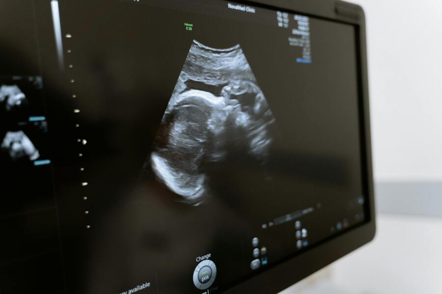
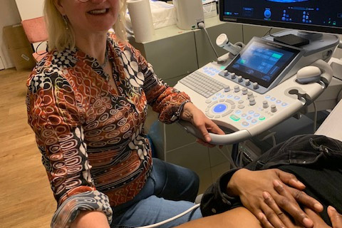
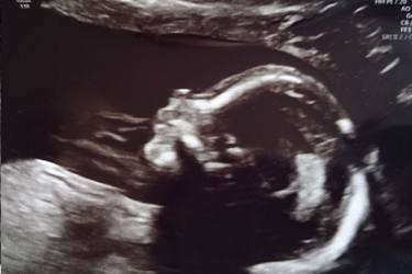
An ultrasound examination that will be offered during pregnancy, but is not mandatory, is the Structural Ultrasound Examination (SEO). This examination is also called the 20-week ultrasound and can be performed from 18 to 22 weeks. Ideally, this ultrasound should be scheduled around 19 weeks of pregnancy. This examination is intended to detect physical abnormalities in the baby, such as spina bifida or skull. In addition, the sonographer will look at all visible structures and organs of the baby during this ultrasound The baby's growth will also be measured and the amniotic fluid and placenta will be examined. The examination is reimbursed by the health insurer. During the first check-up, the midwife will tell you more about this ultrasound examination and this topic will also be discussed during the second check-up. For more information, explanations and video pns ... you can also look at the RIVM website and read the leaflet.

video pns

kun je ook op de website van het RIVM kijken en de folder lezen.

Biometric Ultrasound

If you have an increased risk of having a small or too large baby, we will arrange additional growth ultrasounds (biometrics). During a growth ultrasound we measure the size of the head, the waist circumference and the leg length, this combination gives an estimated weight. We also check the amount of amniotic fluid and look at the position of the baby. Often these are several ultrasounds with an interval of two weeks. With us, everyone gets standard growth ultrasounds at 30 and 34 weeks of pregnancy. It may be that we need more ultrasounds. We always discuss this with you.

Eerste trimester SEO

The 13-week ultrasound is a medical examination for physical abnormalities in the baby. The 13-week ultrasound is very similar to the 20-week ultrasound. In both examinations, an ultrasound technician uses an ultrasound machine to check whether the baby has any physical abnormalities. For example, the sonographer looks at the skull, heart, abdomen, arms and legs and spine during a 13-week ultrasound. The sonographer also looks at the placenta and whether the amniotic fluid is normal. The baby is smaller and less developed at around 13 weeks than at the 20-week ultrasound. Many deviations are not yet visible. But some, often serious abnormalities do. These can be so severe that the baby dies during or shortly after pregnancy. The purpose of the 13-week ultrasound is to detect these serious abnormalities at an early stage. The sonographer can't see everything. If she doesn't see any abnormalities, this is no guarantee that the baby is healthy. Examples of possible physical abnormalities: 

  • Open skull 
  • Open back 
  • Severe abnormalities of the brain 
  • Fracture or large hole in the abdominal wall 
  • Severe facial deformities 
  • Heart defects Abnormalities of arms or legs 

You can only opt for the 13-week ultrasound if you participate in the scientific IMITAS study. The aim of the IMITAS study is to investigate whether the advantages of the 13-week ultrasound outweigh its disadvantages. Click here to read more about the IMITAS study. The research will be reimbursed. During the first check-up, the midwife will tell you more about this ultrasound examination and, if desired, give you more information about it. You will be counselled whether you want to participate in the study For more information, visit the RIVM website RIVM,, watch the video  video and read the leaflet folder


More information about the IMITAS study here 

Placental localisation

Usually as a follow-up to the SEO, the examination is repeated at 34 weeks for a low-lying placenta (placenta praevia). In our practice, growth is then determined as standard. If the placenta is on the back wall, we would also like to check internally whether the placenta is not too close to the cervix.

Gender ultrasound/fun ultrasound

If you want to look at the baby more often, it is also possible to have a fun ultrasound made with us. This is not covered by your health insurance and costs 40 euros. From about 14 weeks, a gender ultrasound is possible. During this ultrasound it is possible to see your baby and you will find out whether it is a boy or a girl. You can let us know if you would prefer that write down the gender and put it in an envelope for you.


Vitality ultrasound at 7-8 weken

An early ultrasound around 7-8 weeks of pregnancy, the sonographer checks whether the fertilized egg has implanted in the right place in the uterus (not ectopic) and whether the heart is beating. Then we check whether there are multiple births. Ultrasound can be done in two ways, externally through the abdomen or internally through the vagina. For ultrasounds before 10 weeks, we prefer an internal ultrasound. The early ultrasound is best seen with an internal vaginal ultrasound. That is why we advise you to urinate prior to this examination. Even in a later period, it can be decided to switch to an internal ultrasound if the external ultrasound does not give good image quality.

CRL/dating scan at 10-12 weken


During the ultrasound, the midwife or sonographer will measure the baby. This measurement is the CRL measurement, which is an abbreviation of the Crown-Rump Length. This measurement can be used to determine exactly how many weeks (and days) you are pregnant. This is because all babies grow at almost the same rate during the first 15 weeks. After that, there will be more individual growth differences. If the final due date after measuring the CRL measurement differs from the date calculated from the first day of your period, the ultrasound date will always be used and the due date will be adjusted.